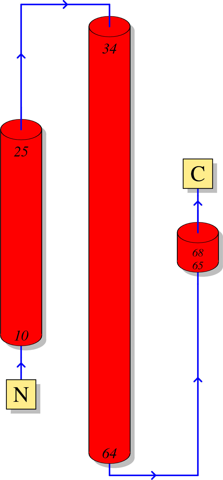
Function of HIV Rev
Rev is an accessory protein which controls the nuclear export of unspliced and partially spliced viral RNAs from nucleus to cytoplasm [1].
Rev multimers bind to the stem-loop structure of Rev response element (RRE) in the env coding region of viral RNA, forming a large oligomeric ribonucleoprotein (RNP) [2].
RNP complex interacts with the human export factor CRM1 (exportin 1 or Xpo1) and shuttles through the nuclear pore complex (NPC) from nucleus to cytoplasm.
Rev activity exerts a strong influence on HIV-1 RNA transport, translation and packaging [3].
Rev is not incorporated in the viral particles [4].
Reference
Fernandes J, Jayaraman B, Frankel A: The HIV-1 Rev response element: an RNA scaffold that directs the cooperative assembly of a homo-oligomeric ribonucleoprotein complex. RNA biology 2012, 9(1):6-11.(Download Article)
Xue B, Mizianty MJ, Kurgan L, Uversky VN: Protein intrinsic disorder as a flexible armor and a weapon of HIV-1. Cellular and molecular life sciences : CMLS 2012, 69(8):1211-1259.(Download Article)
Jeang K-T: Multi-Faceted Post-Transcriptional Functions of HIV-1 Rev. Biology 2012, 1(2):165-174.(Download Article)
Giroud C, Chazal N, Briant L: Cellular kinases incorporated into HIV-1 particles: passive or active passengers? Retrovirology 2011, 8:71.(Download Article)
Sequence
(1) Reference sequence for HIV-1 Rev
  1       10         20         30         40         50
  |        |          |          |          |          |
  MAGRSGDSDE ELIRTVRLIK LLYQSNPPPN PEGTRQARRN RRRRWRERQR
  51      60         70         80         90         100
  |        |          |          |          |          |
  QIHSISERIL GTYLGRSAEP VPLQLPPLER LTLDCNEDCG TSGTQGVGSP
  101    110     116
  |        |      |
  QILVESPTVL ESGTKE
(2) Reference sequence for HIV-2 and SIV Rev
  1       10         20         30         40         50
  |        |          |          |          |          |
  MSNHEREEEL RKRLRLIHLL HQTNPYPTGP GTANQRRQRK RRWRRRWQQL
  51      60         70         80         90         100
  |        |          |          |          |          |
  LALADRIYSF PDPPTDTPLD LAIQQLQNLA IESIPDPPTN TPEALCDPTE
  101  107
  |     |
  DSRSPQD
(3) Coloring scheme for above amino acids
Amino acids with hydrophobic side chains (normally buried inside the protein core):
A - Ala - Alanine
I - Ile - Isoleucine
L - Leu - Leucine
M - Met - Methionine
V - Val - Valine
Amino acids with polar uncharged side chains (may participate in hydrogen bonds):
N - Asn - Asparagine
Q - Gln - Glutamine
S - Ser - Serine
T - Thr - Threonine
Amino acids with positive charged side chains:
H - His - Histidine
K - Lys - Lysine
R - Arg - Arginine
Amino acids with negative charged side chains:
D - Asp - Aspartic acid
E - Glu - Glutamic acid
Amino acids with aromatic side chains:
F - Phe - Phenylalanine
Y - Tyr - Tyrosine
W - Trp - Tryptophan
Cysteine: C - Cys - Cysteine
Glycine: G - Gly - Glycine
Proline: P - Pro - Proline
Amino acid variations at HIV-1 Rev
Here, we visualize the prevalence of amino acid variations at the HIV-1 Rev from HIV-1 subtype B.
Protocal of our sequence collection
For HIV-1 subtype B, one sequence per patient was extracted from HIV Los Alamos database (www.hiv.lanl.gov/).
We removed misclassified sequences or sequences with hypermutations, stop codons, ambiguous nucleotides, which were described in our article [1].
We removed sequences conferred partial or full resistance to any of the Rev inhibitors, RT inhibitors and Rev inhibitors using HIVdb V6.0 .
Visualization
Our sequence dataset of HIV-1 subtype B Rev included 4725 sequences. In the following picture, HXB2 indices of individual proteins are shown on top of the colored bars. A consensus amino acid at each position is shown beneath the colored bar. Natural variations are shown below the consensus amino acids; proportions (%) are colored red if they were more than 5%; blue otherwise.
HIV-1 protein interaction patterns.
Please cite our article:
Guangdi Li, Supinya Piampongsant, Nuno Rodrigues Faria, Arnout Voet, Andrea-Clemencia Pineda-Peña, Ricardo Khouri, Philippe Lemey, Anne-Mieke Vandamme, Kristof Theys. An integrated map of HIV genome-wide variation from a population perspective. Retrovirology 12, 18, doi:10.1186/s12977-015-0148-6 (2015). [PDF] [PubMed Link]


