Function of HIV Gag
- HIV Gag is a structural protein encoded by the gag gene, which provides the basic infrastructure of HIV particles [1].
- Gag in the plasma membrane can recruit host factors (e.g. TIP47) into nascent HIV particles [2].
- Many inhibitors have been developed to target drug binding sites in HIV-1 Gag [3].
Reference
- Bell, N. M. & Lever, A. M. HIV Gag polyprotein: processing and early viral particle assembly. Trends in microbiology 21, 136-144, doi:10.1016/j.tim.2012.11.006 (2013). [PubMed Link]
- Waheed, A. A. & Freed, E. O. HIV type 1 Gag as a target for antiviral therapy. AIDS research and human retroviruses 28, 54-75, doi:10.1089/AID.2011.0230 (2012). [PubMed Link]
- Guangdi Li, Jens Verheyen, Soo-Yon Rhee, Arnout Voet, Anne-Mieke Vandamme, Kristof Theys. Functional conservation of HIV-1 Gag: implications for rational drug design. Retrovirology. 10:126. doi: 10.1186/1742-4690-10-126 (2013). [PubMed Link]
Sequence
(1) Reference sequence for HIV-1 Gag
  1       10         20         30         40         50
  |        |          |          |          |          |
  MGARASVLSG GELDRWEKIR LRPGGKKKYK LKHIVWASRE LERFAVNPGL
  51      60         70         80         90         100
  |        |          |          |          |          |
  LETSEGCRQI LGQLQPSLQT GSEELRSLYN TVATLYCVHQ RIEIKDTKEA
  101    110        120        130        140        150
  |        |          |          |          |          |
  LDKIEEEQNK SKKKAQQAAA DTGHSNQVSQ NYPIVQNIQG QMVHQAISPR
  151    160        170        180        190        200
  |        |          |          |          |          |
  TLNAWVKVVE EKAFSPEVIP MFSALSEGAT PQDLNTMLNT VGGHQAAMQM
  201    210        220        230        240        250
  |        |          |          |          |          |
  LKETINEEAA EWDRVHPVHA GPIAPGQMRE PRGSDIAGTT STLQEQIGWM
  251    260        270        280        290        300
  |        |          |          |          |          |
  TNNPPIPVGE IYKRWIILGL NKIVRMYSPT SILDIRQGPK EPFRDYVDRF
  301    310        320        330        340        350
  |        |          |          |          |          |
  YKTLRAEQAS QEVKNWMTET LLVQNANPDC KTILKALGPA ATLEEMMTAC
  351    360        370        380        390        400
  |        |          |          |          |          |
  QGVGGPGHKA RVLAEAMSQV TNSATIMMQR GNFRNQRKIV KCFNCGKEGH
  401    410        420        430        440        450
  |        |          |          |          |          |
  TARNCRAPRK KGCWKCGKEG HQMKDCTERQ ANFLGKIWPS YKGRPGNFLQ
  451    460        470        480        490        500
  |        |          |          |          |          |
  SRPEPTAPPE ESFRSGVETT TPPQKQEPID KELYPLTSLR SLFGNDPSSQ
(2) Reference sequence for HIV-2 and SIV Gag
  1       10         20         30         40         50
  |        |          |          |          |          |
  MGVRNSVLSG KKADELEKIR LRPNGKKKYM LKHVVWAANE LDRFGLAESL
  51      60         70         80         90         100
  |        |          |          |          |          |
  LENKEGCQKI LSVLAPLVPT GSENLKSLYN TVCVIWCIHA EEKVKHTEEA
  101    110        120        130        140        150
  |        |          |          |          |          |
  KQIVQRHLVV ETGTTETMPK TSRPTAPSSG RGGNYPVQQI GGNYVHLPLS
  151    160        170        180        190        200
  |        |          |          |          |          |
  PRTLNAWVKL IEEKKFGAEV VPGFQALSEG CTPYDINQML NCVGDHQAAM
  201    210        220        230        240        250
  |        |          |          |          |          |
  QIIRDIINEE AADWDLQHPQ PAPQQGQLRE PSGSDIAGTT SSVDEQIQWM
  251    260        270        280        290        300
  |        |          |          |          |          |
  YRQQNPIPVG NIYRRWIQLG LQKCVRMYNP TNILDVKQGP KEPFQSYVDR
  301    310        320        330        340        350
  |        |          |          |          |          |
  FYKSLRAEQT DAAVKNWMTQ TLLIQNANPD CKLVLKGLGV NPTLEEMLTA
  351    360        370        380        390        400
  |        |          |          |          |          |
  CQGVGGPGQK ARLMAEALKE ALAPVPIPFA AAQQRGPRKP IKCWNCGKEG
  401    410        420        430        440        450
  |        |          |          |          |          |
  HSARQCRAPR RQGCWKCGKM DHVMAKCPDR QAGFLGLGPW GKKPRNFPMA
  451    460        470        480        490        500
  |        |          |          |          |          |
  QVHQGLMPTA PPEDPAVDLL KNYMQLGKQQ REKQRESREK PYKEVTEDLL
  501    510
  |        |
  HLNSLFGGDQ
(3) Coloring scheme for above amino acids
Amino acids with hydrophobic side chains (normally buried inside the protein core):
A - Ala - Alanine
I - Ile - Isoleucine
L - Leu - Leucine
M - Met - Methionine
V - Val - Valine
Amino acids with polar uncharged side chains (may participate in hydrogen bonds):
N - Asn - Asparagine
Q - Gln - Glutamine
S - Ser - Serine
T - Thr - Threonine
Amino acids with positive charged side chains:
H - His - Histidine
K - Lys - Lysine
R - Arg - Arginine
Amino acids with negative charged side chains:
D - Asp - Aspartic acid
E - Glu - Glutamic acid
Amino acids with aromatic side chains:
F - Phe - Phenylalanine
Y - Tyr - Tyrosine
W - Trp - Tryptophan
Cysteine: C - Cys - Cysteine
Glycine: G - Gly - Glycine
Proline: P - Pro - Proline
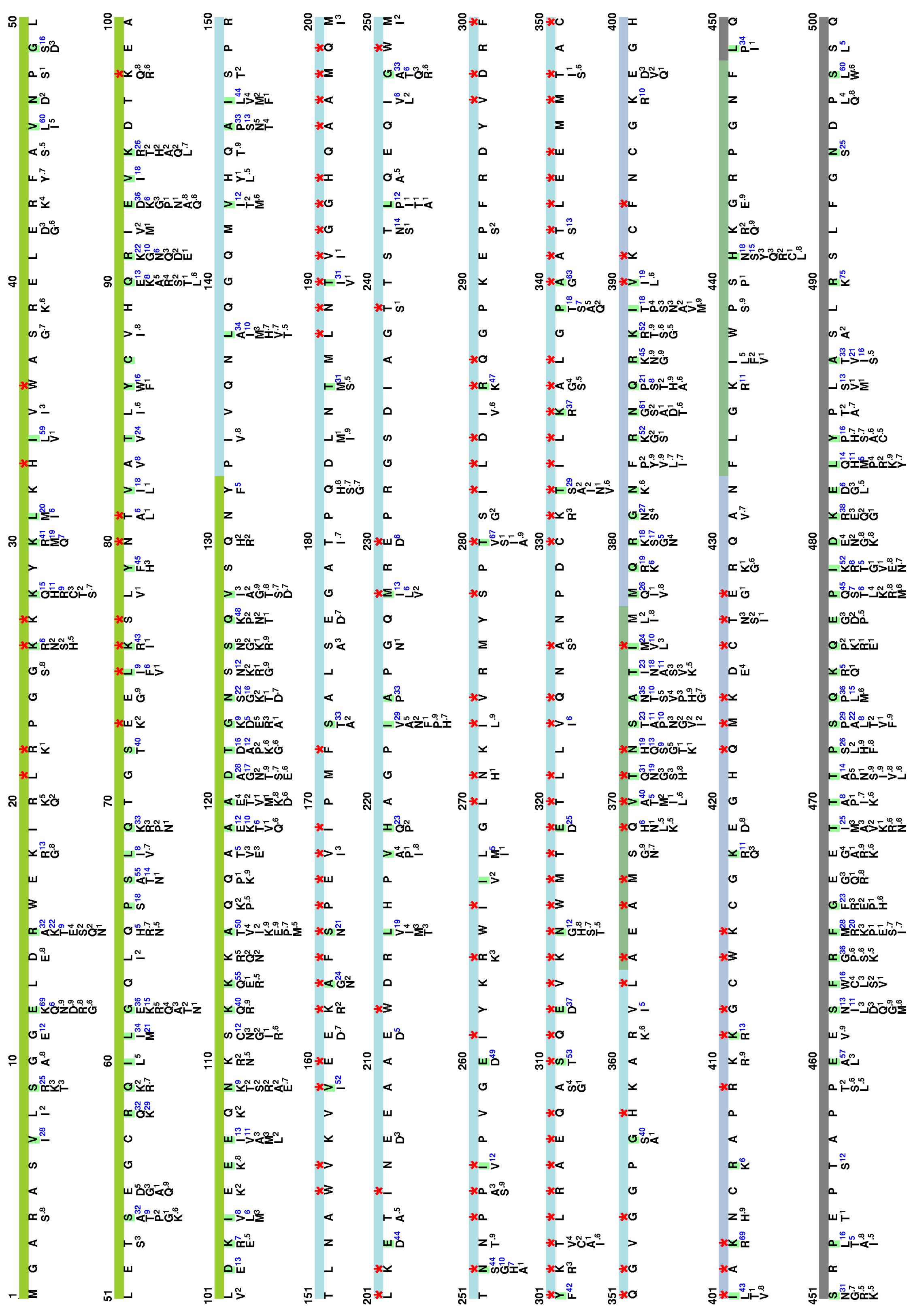
Amino acid variations at HIV-1 Gag
Here, we visualize the prevalence of amino acid variations at the HIV-1 Gag from HIV-1 group M.
Protocal of our sequence collection
Detailed protocals have been described in our publication:
Guangdi Li, Jens Verheyen, Soo-Yon Rhee, Arnout Voet, Anne-Mieke Vandamme, Kristof Theys. Functional conservation of HIV-1 Gag: implications for rational drug design. Retrovirology. 10:126. doi: 10.1186/1742-4690-10-126 (2013). [PubMed Link]
Visualization of natural polymorphisms in Gag
Please cite our article:
Guangdi Li, Jens Verheyen, Soo-Yon Rhee, Arnout Voet, Anne-Mieke Vandamme, Kristof Theys. Functional conservation of HIV-1 Gag: implications for rational drug design. Retrovirology. 10:126. doi: 10.1186/1742-4690-10-126 (2013). [PubMed Link]
Localization
(1) Coding region of Gag at the HIV genome

(2) Localization of Gag during the HIV-1 life cycle
Here, we visualize the localization of Gag during the viral life cycle. Red stars indicate the appearance of HIV-1 Gag.

Anti-HIV inhibitor
(1) Drug binding pocket of HIV-1 Gag
Here, we visualize the drug binding pocket of HIV-1 Gag
