Function of HIV Nucleocapsid
Nucleocapsid is a structural protein encoded by the gag gene [1-4].
To prevent viral RNA from nucleases, Nucleocapsid binds with the genomic viral RNA during viral packaging and coats the genomic RNA within viral core [5].
Nucleocapsid can also bind to host proteins such as the ESCRT-associated protein ALIX to promote viral budding [6].
Served as an RNA chaperone, nucleocapsid enhances nucleic acid-dependent steps in the HIV life cycle. For instance, it promotes the DNA strand exchange reactions during reverse transcription and stimulates viral integration during viral integration [7].
Reference
Mougel M, Houzet L, Darlix JL: When is it time for reverse transcription to start and go? Retrovirology 2009, 6:24.(Download Article)
Darlix JL, Godet J, Ivanyi-Nagy R, Fosse P, Mauffret O, Mely Y: Flexible nature and specific functions of the HIV-1 nucleocapsid protein. J Mol Biol 2011, 410:565-581.(Download Article)
Thomas JA, Gorelick RJ: Nucleocapsid protein function in early infection processes. Virus Res 2008, 134:39-63. (Download Article)
Bell NM, Lever AM: HIV Gag polyprotein: processing and early viral particle assembly. Trends Microbiol 2013, 21:136-144.(Download Article)
Didierlaurent L, Racine PJ, Houzet L, Chamontin C, Berkhout B, Mougel M: Role of HIV-1 RNA and protein determinants for the selective packaging of spliced and unspliced viral RNA and host U6 and 7SL RNA in virus particles. Nucleic Acids Res 2011, 39:8915-8927.(Download Article)
Sette P, Dussupt V, Bouamr F: Identification of the HIV-1 NC binding interface in Alix Bro1 reveals a role for RNA. J Virol 2012, 86:11608-11615.(Download Article)
Xue B, Mizianty MJ, Kurgan L, Uversky VN: Protein intrinsic disorder as a flexible armor and a weapon of HIV-1. Cell Mol Life Sci 2012, 69:1211-1259.(Download Article)
Sequence
(1) Reference sequence for HIV-1 Nucleocapsid
  1       10         20         30         40         50
  |        |          |          |          |          |
  MQRGNFRNQR KIVKCFNCGK EGHTARNCRA PRKKGCWKCG KEGHQMKDCT
  51
  |
  ERQAN
(2) Reference sequence for HIV-2 and SIV Nucleocapsid
  1       10         20         30         40         50
  |        |          |          |          |          |
  AQQRGPRKPI KCWNCGKEGH SARQCRAPRR QGCWKCGKMD HVMAKCPDRQ
  51
  |
  AG
(3) Coloring scheme for above amino acids
Amino acids with hydrophobic side chains (normally buried inside the protein core):
A - Ala - Alanine
I - Ile - Isoleucine
L - Leu - Leucine
M - Met - Methionine
V - Val - Valine
Amino acids with polar uncharged side chains (may participate in hydrogen bonds):
N - Asn - Asparagine
Q - Gln - Glutamine
S - Ser - Serine
T - Thr - Threonine
Amino acids with positive charged side chains:
H - His - Histidine
K - Lys - Lysine
R - Arg - Arginine
Amino acids with negative charged side chains:
D - Asp - Aspartic acid
E - Glu - Glutamic acid
Amino acids with aromatic side chains:
F - Phe - Phenylalanine
Y - Tyr - Tyrosine
W - Trp - Tryptophan
Cysteine: C - Cys - Cysteine
Glycine: G - Gly - Glycine
Proline: P - Pro - Proline
Amino acid variations at HIV-1 Nucleocapsid
Here, we visualize the prevalence of amino acid variations at the HIV-1 Nucleocapsid from HIV-1 subtype B.
Protocal of our sequence collection
For HIV-1 subtype B, one sequence per patient was extracted from HIV Los Alamos database (www.hiv.lanl.gov/).
We removed misclassified sequences or sequences with hypermutations, stop codons, ambiguous nucleotides, which were described in our article [1].
We removed sequences conferred partial or full resistance to any of the protease inhibitors, RT inhibitors and integrase inhibitors using HIVdb V6.0 .
Visualization
Our sequence dataset of HIV-1 subtype B Nucleocapsid included 4725 sequences. In the following picture, HXB2 indices of individual proteins are shown on top of the colored bars. A consensus amino acid at each position is shown beneath the colored bar. Natural variations are shown below the consensus amino acids; proportions (%) are colored red if they were more than 5%; blue otherwise.
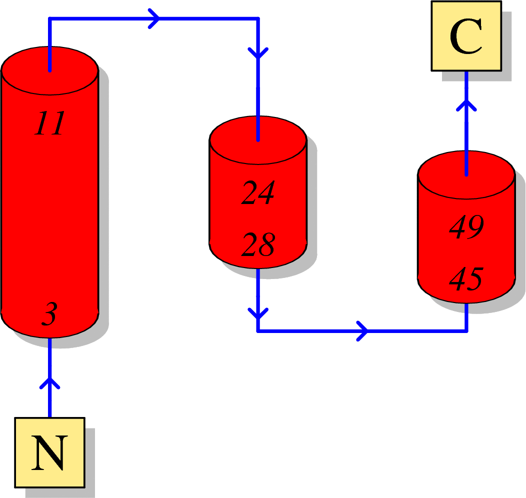
HIV-1 protein interaction patterns.

Please cite our article:
Guangdi Li, Supinya Piampongsant, Nuno Rodrigues Faria, Arnout Voet, Andrea-Clemencia Pineda-Peña, Ricardo Khouri, Philippe Lemey, Anne-Mieke Vandamme, Kristof Theys. An integrated map of HIV genome-wide variation from a population perspective. Retrovirology 12, 18, doi:10.1186/s12977-015-0148-6 (2015). [PDF] [PubMed Link]
Structure
(1) Secondary structure of HIV-1 Nucleocapsid
Here, we visualize the secondary structure of HIV-1 Nucleocapsid using PDBSum (PDB code: 1A1T)
(2) Tertiary structure of HIV-1 Nucleocapsid
Here, we provide a structure movie of Nucleocapsid using PyMOL V1.7 (PDB code: 1A1T). Alpha-helix and beta-strand secondary structures are demonstrated by red .
Localization
(1) Coding region of Nucleocapsid at the HIV genome

(2) Localization of Nucleocapsid during the HIV-1 life cycle
Here, we visualize the localization of Nucleocapsid during the viral life cycle. Red stars indicate the appearance of HIV-1 Nucleocapsid.

Anti-HIV inhibitor
(1) Drug binding pocket of HIV-1 Nucleocapsid
Here, we visualize the drug binding pocket of HIV-1 Nucleocapsid
